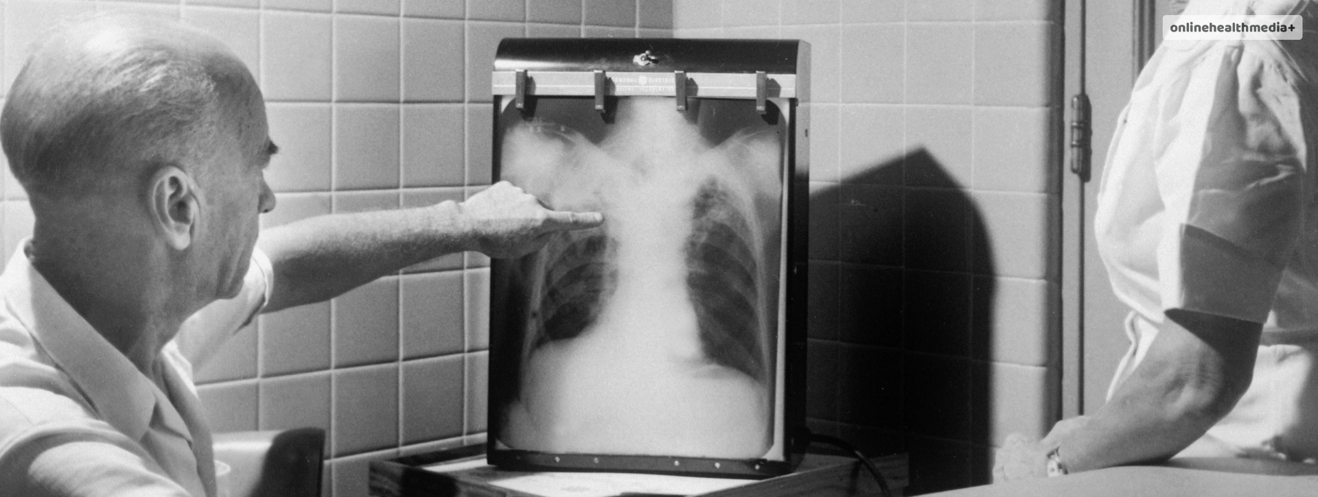
Plueral Effusion 101: What Is It, Causes, Symptoms And More.
Pleural effusion is a condition that is a result of buildup of excess fluids between the layers of the pleura outside the organ. The pleura is a thin membrane that lines the lungs and the inside of the cavity.
Certain conditions can cause the buildup of fluids inside the lungs. A small amount of fluid is always present within the pleura.
When the amount of fluid increases, it causes various issues- these may range from breathing difficulty to the fluid leaking into other organs.
The following article discusses the
Note:
The information in this article does not substitute professional medical advice, diagnosis or treatment. All images and text present are for general information purpose only.
Contents
Pleural Effusion Causes

There can be many causes of pleural effusion. The excess fluid can be protein-rich or protein-poor. Exudative fluid refers to the protein-rich fluid, whereas, transudative fluid refers to the protein-poor or the watery fluid that .
The following list provides an idea of the possible causes of this condition:
- Infection- Bacterial infection may lead to the buildup of transudative pleural effusion, which is the watery fluid that is present.
- Other conditions that cause the buildup of transudative fluid are cirrhosis, or after a major surgery such as open heart surgery. It can also occur due to pulmonary embolism.
- The causes of exudative fluids can also be either cancer, pneumonia, kidney disease or pulmonary embolism and inflammatory diseases.
There can also be other causes of pleural effusion such as:
- Autoimmune disease
- Tuberculosis
- Chylothorax
- Bleeding
- Exposure to asbestos
- Due to the presence of a benign ovarian tumor
- Ovarian hyper-stimulation syndrome
- Chest and abdominal infections which may also be rare
Symptoms of pleural effusion

Despite being a condition that does not really have physical manifestations. However, the following symptoms can help you understand if you have a buildup of excess fluid:
- Chest pain– especially which is prominent during deep breaths
- Shortness of breath
- Fever
- Cough
Additionally, you can experience these symptoms when the pleural effusion is large-sized and if there is inflammation.
Pleural Effusion Diagnosis

Common tests that can help diagnose the condition include-
- CT scan of the chest
- Chest X-ray
- Pleural fluid analysis
- Thoracentesis
- Ultrasound of the chest
- Thoracoscopy (when less invasive tests have not been performed.)
These tests help in identifying the amount of pleural fluid that is present within the lungs while also looking for any leaking.
A minimally invasive procedure, the thoracoscopy helps in diagnosing the cause of pleural effusion. A physician may combine the diagnosis of the condition with the treatment, which is when VATS comes in. VATS is video-assisted thoracoscopic surgery.
Pleural Effusion Treatment

The treatment options for the condition include the following:
- The underlying condition causing pleural effusion can help in developing a treatment plan. For example, if the patient is facing difficulty breathing or has a shortness of breath, the professional can opt for draining of the fluid.
- Other treatment options can include diuretics and other medications for heart failure. This is when the cause of pleural effusion is a congestive heart failure or other medical conditions. The effusion when it becomes malignant may require treatment with radiation, chemotherapy or infusion of medication in the chest.
- The pleural effusion that causes respiratory symptoms can be fixed with the help of draining the liquid using a chest tube.
- A sclerosing agent such as tetracycline or talc and doxycycline helps in preventing pleural effusion.
- Sclerosing agent has been successful in 50 per cent of the cases.
Surgery is another option that can help successfully treat the condition.

- VATS is another approach that helps in management of pleural effusion that maybe difficult to drain.
- This along with sclerosing agents can help prevent the recurrence of fluid build up.
- Thoracotomy, also known as open thoracic surgery is a bigger incision in the chest that helps in draining the fluid that is due to an infection.
- The evacuation of the pleural space is with the help of this procedure. It also helps in the removal of the fibrous tissue.
- The presence of a malignant pleural effusion signifies that the condition has progressed to an advanced stage. Its presence is indicative of death. In this case, the management involves palliative care. However, there are low chances of survival benefits.
Palliative care may include taking care of the symptoms and improving the patient’s experience of the condition, especially their uptake of the treatment options.
- The goals of palliative care are relieving the symptoms of the patients. It also helps in the restoration of the patient’s quality of life.
- The intervention should also be affordable for them, as well as be less invasive.
It is worth noting that treatment of the symptoms is dependent on the type of effusion as it helps in understanding how it can be best managed. The Chylous effusions, for example, require surgery for proper drainage.
Guidelines On Pleural Effusion Management
The following list displays the guidelines that help in the proper management of the condition:
- Ultrasound can help in the detection of pleural fluid sequestration.
Bedside ultrasound can help in successfully diagnosing the condition, as well as reduce the risk of pneumothorax during aspiration. - The fluid that is retrieved must be sent for further testing so that the underlying cause can be ascertained.
- The pH of the pleural effusion is also a great way to tell you about the cause of the fluid accumulation. For example, if pH of the sample is less than 7.2, the fluid should be drained.
- The air or local anesthetic should not be added to the sample as it can alter the fluid’s pH.
- When the complete removal of the fluid cannot be done, a CT scan helps in visualization.
- Thoracoscopy can help in diagnosing the malignancy.
Routine bronchoscopy is not done for pleural effusions as it may be dangerous.
Conclusion
This is all on the condition which is known as pleural effusion. The article covers the description of the condition, including the causes, symptoms, its diagnosis and the possible treatment options one can opt for.
The article aims to give you, the valuable reader, a complete rundown of the condition so that they can recognize it easily.
Also read
- Top Electric Toothbrush For Kids.
- The Most Common Types Of Bicycle Accident Injuries.
- Compelling Reasons To Incorporate Shilajit Into Your Diet.


