Thyroid Eye Disease Radiology: Everything You Need to Know.
Thyroid eye disease radiology is an imaging technique that helps visualize the condition. This diagnostic procedure helps study the abnormalities that come up due to thyroid eye disease.
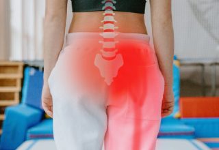
Thyroid eye disease is a complex disorder involving the involvement of the adipocytes, fibroblasts, extra-ocular muscles, orbital vasculature, and optic nerve.
The condition has different clinical presentations, which can range from unilateral to bilateral proptosis. There are also other conditions related to this condition, which will be discussed in the following section.
The condition is also known as TED, whose mention you will see repeatedly through the text. Being related to Graves’ disease, nearly half of the 50% of cases present with thyroid eye disease.
The pathogenesis of the condition involves orbital fibroblasts, which are the target cells. Moreover, the condition also includes the formation of mature adipocytes.
The following article delves deep into the condition, the role of radiology in its diagnosis, and other related information. Read on!
What Is Thyroid Eye Disease?

Nearly 30% of the patients with a diagnosis of Graves’ disease present the signs of Graves’ eye disease, also known as thyroid eye disease.
Most GED patients develop mild symptoms, which may include inflammation or fat expansion. The more severe cases include eye retraction, conjunctival edema, or compression of the optic nerve.
The last condition may lead to compressive optic neuropathy or CON. The severity of the condition is as per the Clinical Activity Score, a type of classification system.
Recent studies demonstrate through MRI findings that the condition’s severity can help in evaluating CON. Moreover, a study also found that mild cases of TED can lead to spontaneous recovery within a couple of years.
The condition may also require medical treatment. However, it is mostly used to limit immunologic damage and may include radiotherapy or immunomodulators.
What Is Thyroid Eye Disease Radiology?

Radiology is an imaging technique that includes diagnosing and treating disease. The determination of a medical condition may include radiology as a tool.
It may also help as a procedure or even as a treatment. For example, it’s used in removing blood clots in an artery or in treating cancer, respectively.
Moreover, with advancements in technology, the use of artificial intelligence in the healthcare sector is all the rage. AI is becoming increasingly finding its way into radiology and healthcare.
The expansion helps in using the strengths of AI such as accuracy, automation, and objectivity. However, this is yet to find its way into diagnosing thyroid eye disease.
Even if you have the question “How to reduce swelling from thyroid eye disease?” radiology will be able to provide an answer.
Radiology in thyroid eye disease is usually used first in the diagnosis stage, where the primary care physician uses it.
The confirmatory diagnosis of the condition can only be received once there is proper visualization of the condition’s severity.
There are three main types of radiology, mainly:
- Diagnostic Radiology
- Interventional radiology
- Radiation oncology
The healthcare professional who is skilled in performing either of the three types of all three is known as a radiologist.
In this scenario, there can be a condition or an injury that a radiologist is diagnosing or treating. This includes either of the following:
- X-rays
- Ultrasound
- Fluoroscopy
- Computed Tomography or CT scan. It is also known as Computerized Axial Tomography CAT scan.
- MRI or Magnetic Resonance Imaging
- Fusion imaging is when two different imaging tests are fused
- Mammography
- Nuclear medicine imaging
Thyroid Eye Disease Radiology: Diagnostic Radiology

This is the branch of radiology that includes the use of imaging equipment such as those listed above for diagnosis.
For example, in conditions such as thyroid eye disease, radiology can be used to scan the affected area. The experts can interpret the images and help the professional arrive at a diagnosis.
They may even use the imaging technique to monitor the body’s response to the treatment being administered.
Interventional radiology helps in guiding a procedure. For example, radiology will find its way through CT or MRI scans in the case of thyroid eye disease.
Radiology helps guide the procedure, such as deciding if surgery must be chosen as an option. The interventional radiologist’s presence may be necessary in treating TED, especially if it involves complications.
Radiation oncology
The involvement of radiation is not just in imaging techniques. Radiation can also be used to treat conditions that do not comply with usual treatment options.
Treatment of cancer, for example, includes radiation. Radiation oncology includes treating non-cancerous as well as cancerous conditions.
Cancer treatment uses radiation to kill the cells or at least inhibit their growth and division. Radiation therapy maximizes the effectiveness of the treatment and minimizes any harm to surrounding healthy cells.
Frequently Asked Questions (FAQs)
1. How can I practice thyroid eye disease radiology?
Thyroid eye disease radiology includes going to medical school during graduation. Then, you can also apply for a license that allows you to practice depending on the area with an Eye Doctor.
It may include becoming either a diagnostic radiologist, interventional radiologist, or radiologist in oncology.
2. How to become a radiologist?
Becoming a radiologist (no matter the type) requires:
- Obtaining pre-medical education at a university or college and graduating with a bachelor’s degree.
- Graduating from an accredited medical school
- Pass the USMLE or the United States Medical Licensing Examination.
- Complete a residency of at least four years.
One or two additional years that focus on a specialization. Such as that you are training in a subspeciality of radiology. Such as:
- Diagnostic Radiology
- Pediatric Radiology
- Radiation oncology
- Interventional radiology
- Nuclear medicine
3. How long will it take for me to become a radiologist?
It can take anywhere from 10 to 13 years to become a radiologist. This includes:
- Undergraduate degree
- Medical school
- Four-year residency
- One- or two-year fellowship in the chosen specialty. It isn’t mandatory; however, most radiologists complete this, too.
Conclusion
Thus, the article on thyroid eye disease radiology concludes here. The article described the condition, what radiology is, and how it relates to it.
The text also includes common questions you may have as a reader and an elaboration on the types of radiology.
Have any more questions for us? Let us know in the comment section below.
Also read
- Dry Eye Mask: A Quick Guide To Dry Eye Therapy.
- Been Diagnosed with Diabetes? 6 Critical Things to Do Next.
- Why Should You Consider Wearing Blue Light Filter Glasses?