What Happens During Stent Placement?
A heart attack is one of the most frightening possibilities of growing older, but that doesn’t mean it’s uncommon. Most people will have heart issues at some point in their lifetime.
Over the years, various treatments have been discovered to help aid in recovery after suffering a heart attack. One of the more common medical treatments is stent placement. You’ve heard the word before but might not know the procedure it refers to.
Read on to learn about getting a coronary angioplasty, and what happens during this surgical procedure. Let’s get into it!
Determining The Exact Location Of The Blockage

Medical imaging is used to determine the exact location of the blockage. This imaging provides a detailed look at a patient’s arteries and blood vessels to assist in precise stent placement. There are several ways to visualize the target vessel such as:
- Computed Tomography or CT scans
- Magnetic Resonance Angiography or MRA scans
- Ultrasound imaging
Additionally, it allows the interventionist to make any necessary adjustments throughout the procedure. By having an accurate picture of the vessel, the isolationist can ensure the stent is placed correctly and minimize the risk of any potential complications.
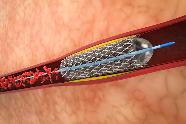
Steps Involved in Stent Insertion

During stent placement, the patient lies on a table and the area of the body to be treated is imaged using an x-ray and real-time ultrasound. A device is then used to insert a tiny tube, or catheter, into the artery to the site of the blockage.
Additionally, the stent is then pushed through the catheter, deployed, and expanded to keep the vessel open. Furthermore, it will remain in place, allowing blood to flow and restoring healthy circulation.
Recovery and Aftercare
The entire process usually takes 1-2 hours and patients typically can go home the same day. To ensure the stent remains in place, it is important to take blood thinners and other medications to prevent blood clots.
Regular check-ups are also recommended to ensure the stent is sleeping properly to maintain maximum blood flow.
Interventional Vascular Radiology
Interventional Vascular Radiology is a minimally invasive treatment option typically used when a patient has a blockage present in the artery. This obstruction can be caused by atherosclerosis, a build-up of plaque in the arteries, often caused by high cholesterol.

The treatment involves the placement of stents to keep the artery open and allow blood to flow freely through it. During the procedure, a radiologist makes a small incision in the artery and then inserts a thin tube guided by an x-ray machine and contrast material.
After the stent is in place, a dye is injected through it to make sure the artery is clear and open. If you’re considering this procedure, read about interventional vascular radiology today and see if it is right for you!
A Guide to Stent Placement Procedure
Stent placement is a relatively safe procedure that is effective in treating narrowed arteries. As always, every individual is different and all treatments should be discussed with a medical professional.
If you are experiencing any chest pain, contact your physician, as they’re best equipped to determine the best course of action for your health.
Did you find this article helpful? Check out the rest of our blog now!
Additional:
- Living With Atrial Fibrillation
- What Does A Chest Compression Feedback Device Monitor Do?
- What Are Blood Cell Disorders? Symptoms,Treatment, And Prevention
- How to Deal with Mental Health Issues Such as Depression, Panic Attacks and Anxiety